The MRI
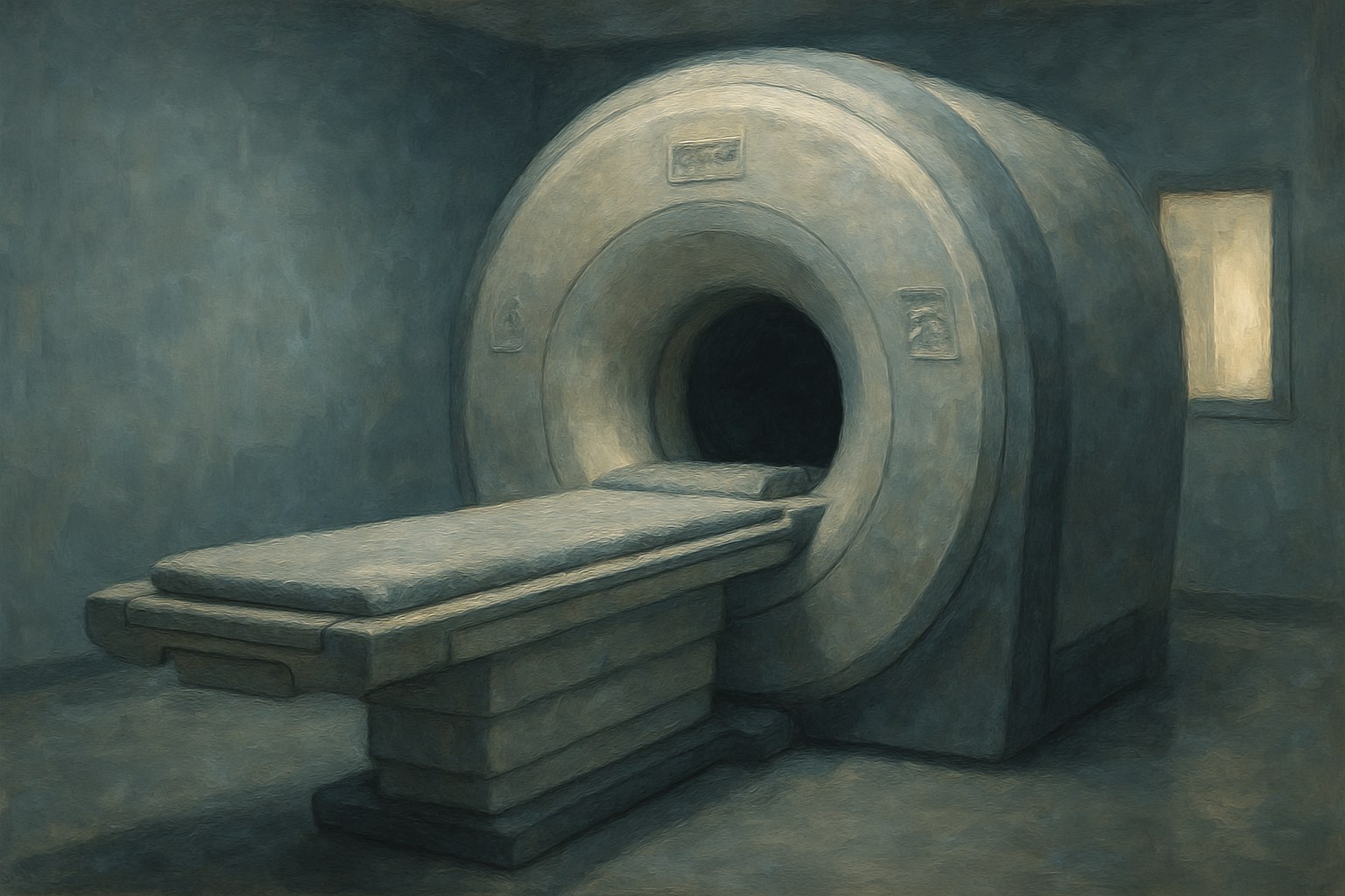
X-rays had shown doctors bone since 1895, and computed tomography added cross-sections in the 1970s, but both worked by firing ionizing radiation through a person and both were poor at soft tissue. The brain, the spinal cord, ligaments, and tumors buried in muscle remained effectively invisible short of cutting. Nuclear magnetic resonance, a laboratory technique for identifying chemical compounds by how their nuclei behave inside a strong magnetic field, had existed since the 1940s. For a quarter century nobody thought of it as a way to look inside a living human being.
In 1971 Raymond Damadian, a physician at what is now SUNY Downstate in Brooklyn, published a paper in Science reporting that cancerous tissue in rats returned measurably different relaxation signals than healthy tissue. That established a medical use, but not a picture. The imaging method itself came from Paul Lauterbur at Stony Brook, who realized in 1973 that superimposing a magnetic gradient across a sample would encode spatial position into the signal, and from Peter Mansfield in Nottingham, whose mathematics made the scans fast enough to be clinically practical.
Damadian built a machine anyway. He and two graduate students spent years winding a superconducting magnet by hand, named the apparatus Indomitable, and on July 3, 1977 produced the first magnetic resonance image of a whole human body — a coarse cross-section of a chest that took nearly five hours to acquire. When the 2003 Nobel Prize in Physiology or Medicine went to Lauterbur and Mansfield, Damadian was left out. He bought full-page newspaper advertisements protesting the omission. The Nobel committee has never explained its reasoning, and the priority dispute has never been resolved.
What the machine delivered was a clear view of the living soft body at no radiation dose at all. Multiple sclerosis lesions, torn knee cartilage, strokes in progress, and tumors deep in the brain became visible to anyone trained to read the images, and exploratory surgery performed simply to find out what was wrong largely disappeared. Functional MRI, developed in the early 1990s, extended the technique to watching blood flow shift as a person thinks. Tens of thousands of scanners now operate worldwide, and the cost of running them is a standing item in every argument about American health spending.
| First medical paper | Raymond Damadian, Science, 1971 |
| Imaging method | Paul Lauterbur, gradients, 1973 |
| Made fast by | Peter Mansfield, Nottingham |
| First body scan | July 3, 1977 — the machine called Indomitable |
| Nobel Prize | 2003, to Lauterbur and Mansfield — Damadian excluded |
| Key advantage | Soft-tissue detail with no ionizing radiation |
| Date | 1971-1977 |
| Location | Brooklyn, New York |